Endoscopic tympanoplasty for chronic suppurative otitis media
Chronic suppurative otitis media is a common otologic disease that occurs due to purulent lesions of the mammilla, periosteum, and bone of the middle ear, resulting in prolonged or paroxysmal pus flow in the ear. If left untreated, it can lead to perforation of the periosteum, hearing loss, and in severe cases, intracranial complications that can lead to death.
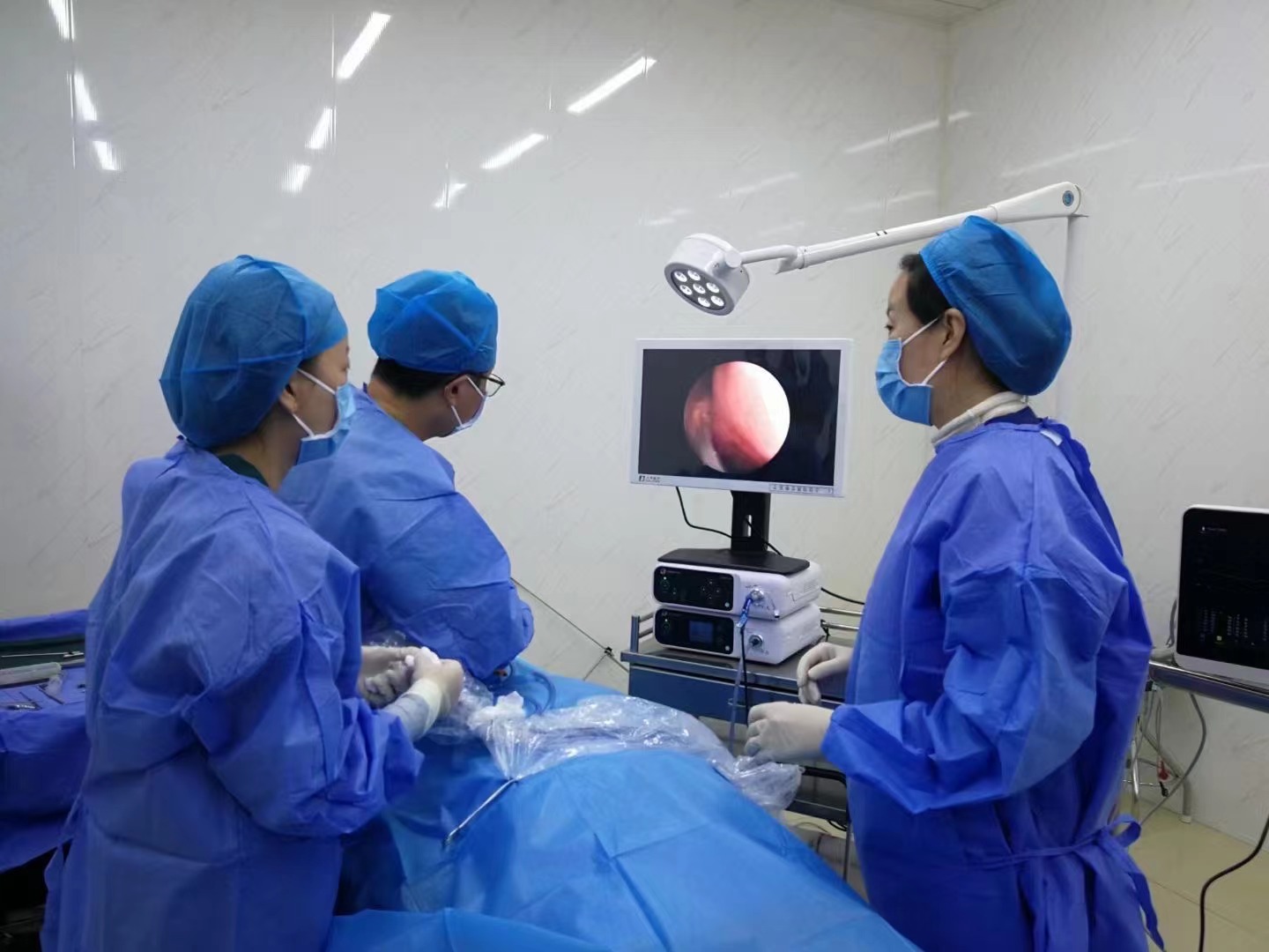
With the development of endoscopic technology, more and more experts are recommending the application of endoscopy in the treatment of chronic suppurative otitis media, which has the advantages of clear vision and less trauma. From the current clinical research, the treatment of chronic suppurative otitis media is mainly carried out through two methods: conservative treatment and surgical treatment, which is usually adopted when the effect of conservative treatment is not satisfactory. In recent years, the use of otoscopy has been increasing. The application of this technology can effectively improve the image clarity, allow thorough observation of the lesioned parts of the inner ear cavity, and make the surgery less invasive. With the aid of an otoscope, the lesion can be observed from an ideal angle, and even in the case of hidden lesions, it can be detected and treated in a timely manner. For patients with narrow external auditory canals or high neural crests, the use of an endoscope can greatly improve the success rate of surgery and lesion identification, reduce the damage to the surrounding normal tissues during surgery, and protect the facial nerve.
However, it is important to note that the lens of the endoscope can easily be contaminated and therefore needs to be withdrawn for cleaning in a timely manner. It is important to be gentle in this process and to choose the appropriate endoscopic treatment method according to the patient's specific situation to ensure efficacy and reduce complications and patient safety. The following points should be noted when using endoscopic surgery: ① Select the appropriate surgical approach according to the patient's ear structure as much as possible, and choose an endoscope with high clarity and brightness as much as possible. (2) Minimize pulling during surgery to avoid damage to other tissues. ③In case of serious bleeding, the bleeding should be stopped and the lens should be cleaned to ensure the surgical field.
