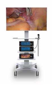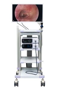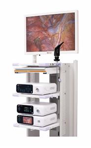
Transnasal endoscopic resection of nasopharyngeal fibroangioma
Through the nasal cavity, nasopharynx and CT examination, it was found that all tumors invaded the affected nasal cavity from the nasopharynx, and 2 cases invaded the sphenoid sinus. According to Chandler's staging method, all of them were stage II. Five patients were treated with general anesthesia. The external carotid artery on the affected side was first ligated, controlled hypotension was performed, and then bipolar electrocoagulation, microwave, etc. were used to conduct extraperiosteal thermal coagulation separation and resection through the nasal cavity under the guidance of nasal endoscope. Norepinephrine gauze was used to tightly fill the wound area to compress the tumor and help stop bleeding. The tumor base was resected subperiosteally, and the size of the resected tumor ranged from 4 cm×3 cm×2 cm to 5 cm×4 cm×3 cm. The surgical bleeding was 200-400 ml. Results All 5 patients were discharged from the hospital 7 days after the operation, and were followed up for 3 to 9 years. There was no recurrence, and the nasopharyngeal cavity was smooth and well epithelialized. Conclusion Nasal endoscopic resection of stage Ⅰ and stage Ⅱ nasopharyngeal fibroangioma is a good minimally innovative method. Before the operation, the supplying artery should be blocked and the technique of controlled hypotension should be used to reduce bleeding. The use of this method should be limited to tumors of stage III and stage IV or above.