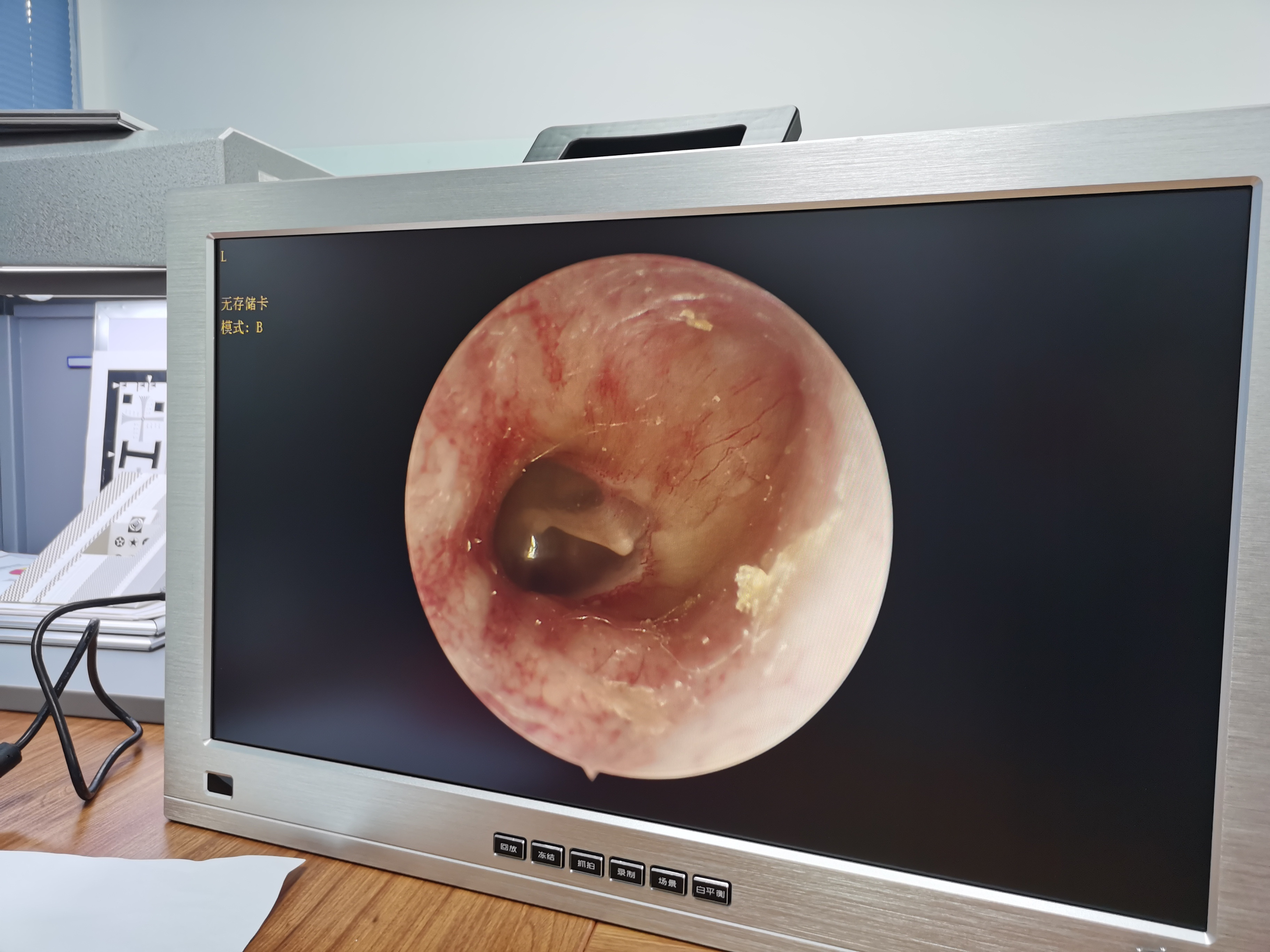
Tragus cartilage-perichondrium repair of tympanic membrane under otoscope and otology microscope in the treatment of perforated tympanic membrane
The clinical data of 142 patients with tympanic membrane perforation who received tympanic membrane repair in our department from May 2015 to January 2016 were divided into ear endoscopic group and microscope group according to the operation method, with 71 cases in each group. Tragus cartilage-perichondrium tympanoplasty and tragus cartilage-perichondrium tympanoplasty under microscope. The operation time, intraoperative blood loss, tympanic membrane healing rate and hearing threshold at 1 month, 3-6 months and 1-2 years after operation were compared between the two groups. Results The operation time of the otoscope group was shorter than that of the microscope group, and the blood loss during operation was less than that of the microscope group, the differences were statistically significant (P<0.05). There was no significant difference in the tympanic membrane healing rate and reperforation rate between the two groups at 1 month, 3-6 months, and 1-2 years after operation (P>0.05). At 1 month and 3 months after operation, the average air conduction threshold and average air-bone conduction difference of the two groups were lower than those before operation, and the difference was statistically significant (P<0.05). There was no statistically significant difference in air-bone difference (P>0.05). During the 1-year follow-up, there were no atrophy of the tympanic membrane, adhesion of the tympanic cavity, and outward migration of the graft. Conclusion The tragus cartilage-perichondrium repair of tympanic membrane perforation under ear endoscope can obtain the same clinical effect as the traditional operation, but the operation time is shorter and the trauma is smaller, which is worthy of clinical promotion.
