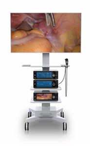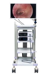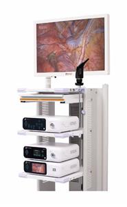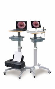
Middle Ear Endoscopy
Author:Drew M Horlbeck,MD
History
Mer, in 1967, was the first to describe use of an endoscope to view the anatomy of the middle ear. He initially performed endoscopy through a tympanic membrane incision on cadavers and on a feline model. Endoscopy in human patients was only attempted through previously existing perforations. Despite his initial success, the endoscope had only limited use as an instrument to photograph the tympanic membrane.
Two decades would pass before Nomura popularized the idea of a surgical myringotomy in an intact drum to permit endoscopic examination of the middle ear structures. Subsequently, direct endoscopy through the middle ear space has been used successfully (1) as an alternate procedure for second-look mastoidectomy, middle ear exploration for perilymphatic fistula, and limited removal of epitympanic cholesteatoma and as an intraoperative aid to visualize the attic, eustachian tube, and sinus tympani.
In 1989, Kimura introduced the concept of endoscopy of the middle ear through the eustachian tube orifice. This technique has not met with the same clinical success as transtympanic endoscopy because of the small image size provided by the scope, difficult orientation, and poor illumination. In a recent study, 25% of endoscopy attempts through the eustachian tube orifice were aborted because of local irritation, bleeding, thick mucus, and/or blocked view by bony spicules. Theoretically, when the difficulties of this technique are overcome, it may provide information pertaining to pathologies in the epitympanum, mesotympanum, and mastoid antrum, and it may allow assessment of ossicular chain mobility. For now, however, the authors find only the transtympanic route for middle ear endoscopy to be a clinically viable technique.
Relevant Anatomy
The ear is composed of external, middle (tympanic) (malleus, incus, and stapes), and inner (labyrinth) (semicircular canals, vestibule, cochlea) portions. The auricle and external acoustic meatus (or external auditory canal) compose the external ear. The external ear functions to collect and amplify sound, which then gets transmitted to the middle ear. The tympanic cavity (middle ear) extends from the tympanic membrane to the oval window and contains the bony conduction elements of the malleus, incus, and stapes. The primary functionality of the middle ear is that of bony conduction of sound via transference of sound waves in the air collected by the auricle to the fluid of the inner ear. The inner ear, also called the labyrinthine cavity, is essentially formed of the membranous labyrinth encased in the bony osseus labyrinth. The labyrinthine cavity functions to conduct sound to the central nervous system as well as to assist in balance.