Functional Endoscopic Sinus Surgery 2
Relevant Anatomy
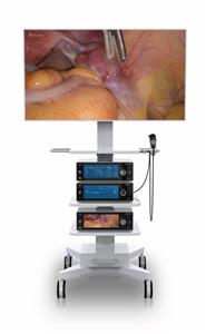
Intimate knowledge and understanding of the anatomy of the lateral nasal wall and the sinuses (see the image below), in conjunction with a careful preoperative review of CT scans, are paramount in the safe and complete performance of endoscopic sinus surgery. The following description of endonasal anatomy is roughly based on the order of dissection during nasal endoscopy and surgery.


Nasal septum and inferior turbinate
Immediately upon entering the nasal cavity, the first structures encountered are the nasal septum and the inferior turbinate. The nasal septum consists of the quadrangular cartilage anteriorly, extending to the perpendicular plate of the ethmoid bone posterosuperiorly and the vomer posteroinferiorly.
Recognizing deflections of the nasal septum preoperatively is important because they may significantly contribute to nasal obstruction and limit endoscopic visualization during surgery. As appropriate, patients with septum deflections may be counseled regarding the need for septoplasty in conjunction with functional endoscopic sinus surgery.
The inferior turbinate extends along the inferior lateral nasal wall posteriorly toward the nasopharynx. In patients with a significant allergic component to their problems, the inferior turbinates may be edematous. These patients may benefit from a turbinate reduction at the same time as the endoscopic sinus surgery. The inferior meatus, where the nasolacrimal duct opens, is located approximately 1 cm beyond the most anterior edge of the inferior turbinate.
Middle turbinate
As the endoscope is further advanced into the nose, the next structure encountered is the middle turbinate. The middle turbinate is a key landmark in endoscopic sinus surgery. It has a vertical component (lying in the sagittal plane, running from posterior to anterior) and a horizontal component (lying in the coronal plane, running from medial to lateral).
Superiorly, the middle turbinate attaches to the skull base at the cribriform plate. As such, care should always be taken when manipulating the middle turbinate.
The horizontal component of the middle turbinate is referred to as the basal (or grand) lamella, and it represents the dividing point between anterior and posterior ethmoid air cells. Posteriorly and inferiorly, the middle turbinate attaches to the lateral nasal wall at the crista ethmoidalis, just anterior to the sphenopalatine foramen.
Uncinate process
The uncinate process is the next key structure to be identified in endoscopic sinus surgery. This L-shaped bone of the lateral nasal wall forms the anterior border of the hiatus semilunaris, or the infundibulum. The infundibulum is the location of the ostiomeatal complex, where the natural ostium of the maxillary sinus opens.
For patients with sinus disease, a patent ostiomeatal complex is critical for improvement of symptoms. Anteriorly, the uncinate process attaches to the lacrimal bone, and inferiorly, the uncinate process attaches to the ethmoidal process of the inferior turbinate.
Natural maxillary ostium
Once the uncinate process is removed, the natural maxillary ostium can be seen, typically just posterior to the uncinate process, roughly one third of the distance along the middle turbinate from its anterior edge. It lies at approximately the level of the inferior border of the middle turbinate, superior to the inferior turbinate.
The natural maxillary ostium is the destination for the mucociliary flow within the maxillary sinus. Therefore, for optimal results, the surgically enlarged maxillary antrostomy must include the natural ostium. In fact, failure to include the maxillary ostium in endoscopic surgical antrostomy is one of the key patterns of failure in functional endoscopic sinus surgery.
The maxillary sinus, approximately 14-15 mL in volume, is bordered superiorly by the inferior orbital wall, medially by the lateral nasal wall, and inferiorly by the alveolar portion of the maxillary bone.
Ethmoid bulla
The next structure to be encountered is the ethmoid bulla, which is one of the most constant anterior ethmoidal air cells. It is just beyond the natural ostium of the maxillary sinus and forms the posterior border of the hiatus semilunaris.
The lateral extent of the bulla is the lamina papyracea. Superiorly, the ethmoid bulla may extend all the way to the ethmoid roof (the skull base). Alternatively, a suprabullar recess may exist above the roof of the bulla. A careful preoperative review of the patient's CT scan clarifies this relationship.
Ethmoid sinus
The ethmoid sinus consists of a variable number (typically 7-15) of air cells. The most lateral border of these air cells is the lamina papyracea, and the most superior border of these cells is the skull base. Supraorbital ethmoid cells may be present. A review of the patient's CT scan alerts the surgeon to these variations.
The basal lamella of the middle turbinate separates the anterior ethmoid cells from the posterior ethmoid cells. Anterior ethmoid cells drain to the middle meatus, and the posterior cells drain into the superior meatus.
Sphenoid sinus
Exenteration of the posterior ethmoid cells exposes the face of the sphenoid. The sphenoid sinus is the most posterior of the paranasal sinuses, sitting just superior to the nasopharynx and just anterior and inferior to the sella turcica. The anterior face of the sphenoid sits approximately 7 cm from the nasal sill on a 30° axis from the horizontal.
Several important structures are related to the sphenoid sinus. The internal carotid artery is typically the most posterior and medial impression seen within the sphenoid sinus. In approximately 7% of cases, the bone is dehiscent.
The optic nerve and its bony encasement produce an anterosuperior indentation within the roof of the sphenoid sinus. In 4% of cases, the bone surrounding the optic nerve is dehiscent. Therefore, controlled opening of the sphenoid sinus, typically at its natural ostium, is critical for a safe outcome.
The location of the natural ostium of the sphenoid sinus is variable. In approximately 60% of people, the ostium is located medial to the superior turbinate, and in 40%, it is located lateral to the superior turbinate.
Frontal recess
The frontal recess, or the frontal sinus outflow tract, is the tract that leads from the frontal sinus into the nasal cavity. Often, the ethmoid bulla is the posterior border of the frontal sinus outflow tract.
Anteriorly, the frontal sinus outflow tract is bordered by the uncinate process or the agger nasi cells (frontal anterior ethmoid air cells). If any of these cells are enlarged or if scarring is present from a previous surgery, resultant outflow tract obstruction, leading to frontal sinusitis, may occur. Typically, the medial wall of the frontal recess is formed by the lamina papyracea.