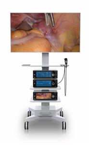
Endoscopic Mucosal Resection (EMR) and Endoscopic Submucosal Dissection (ESD)
Background
Endoscopic mucosal resection
Endoscopic mucosal resection (EMR) is a technique used for the staging and treatment of superficial neoplasms of the gastrointestinal (GI) tract. This technique was first developed in Japan for the treatment of early gatric cancer(EGC) and has since spread in use throughout the world for various indications, including dysplastic Barrett mucosa and sessile colonic neoplasms. The utility of EMR rests in its ability to do the following:
- Provide accurate histologic staging of superficial GI neoplasms
- Provide a minimally invasive technique for removal of superficial malignancies
Several variations of EMR are currently used, including injection-assisted, cap-assisted, and ligation-assisted techniques. All adhere to the basic principles of identification and demarcation of the lesion, submucosal injection to lift the lesion, and endoscopic snare resection. By virtue of its overall safety and efficacy in appropriately selected patient populations, EMR has become firmly integrated into the diagnostic and treatment algorithms of superficial GI malignancies.
Some authors have also studied the use of EMR in a procedure known as antireflux mucosectomy (ARMS) to treat refractory gastroesophageal reflux disease (GERD).
Endoscopic submucosal dissection
Endoscopic submucosal dissection (ESD) was developed to resect larger tumors and aid in achieving higher rates of en-bloc resection than would be possible with EMR. The main goal of ESD is to achieve an R0 resection. In the United States, ESD is primarily performed in select centers by specialized endoscopists experienced in this technique. ESD is generally indicated for the following :
- Tumors that are diagnosed as carcinomas with intramucosal to superficial submucosal invasion
- Lesions with submucosal fibrosis that cannot be removed by EMR even if smaller than 20 mm
- Cases where a snare is unlikely to enable a successful en-bloc resection with EMR
- Removal of large polyps, early colorectal cancer, and those lesions that cannot be accessed transanally in patients who wish to avoid major surgical resection
Endoscopic and ultrasonographic characterization of lesions
Several classification systems for the staging of early GI cancers that may aid in the prediction of lymph node metastases have been developed. Much of this work was pioneered by Japanese gastroenterologists for the staging and treatment of EGC. The Japanese Society of Gastroenterology (JSGE), working from large databases of EGC resections, classified lesions according to their endoscopic features and the implied risk of mural invasion.
The subsequent Paris classification, developed in 2002 at an international consensus meeting, echoed the structure of the JSGE system. In this classification, superficial (type 0) lesions are divided into polypoid (0-I) and nonpolypoid categories (0-II), which are further subcategorized as pedunculated (0-Ip), sessile (0-Is), slightly elevated (0-IIa), flat (0-IIb), slightly depressed (0-IIc), or excavated (0-III).
In the Vienna classification, lesions are divided into two broad categories, noninvasive (low-grade dysplasia, high-grade dysplasia [HGD]) and invasive (intramucosal cancer, cancer that infiltrates the submucosa).
The mucosal layer is divided into upper, middle, and lower layers: m1 (epithelium), m2 (lamina propria), and m3 (muscularis mucosae). The submucosa is similarly divided into three layers: sm1, sm2, and sm3. Submucosal tumor involvement of 500 μm or less below the muscularis mucosae is characterized as sm1 (superficial) disease, and involvement past 500 μm is categorized as sm2-3 (deep) disease. The sm1 layer is further divided into sublayers a, b, and c on the basis of the lateral spread within the layer.
High-frequency (≥20 MHz) endoscopic ultrasonography (EUS) produces an image of the mucosal wall comprising nine separate layers differentiated by their echogenicity. By carefully examining the depth of lesion penetration into the mucosal and submucosal layers, one may determine the risk of lymph node metastases with greater precision.
EUS has excellent sensitivity and specificity in accurately diagnosing the tumor depth and lymph node status of esophageal cancer and is considered the most accurate imaging modality currently available. Its accuracy ranges from 75% to 82% for T1 disease, from 88% to 100% for T4 disease, and from 72% to 80% for lymph node involvement.
A study assessing staging with radiologic and pathologic correlation in patients with esophageal cancer with lymph node metastases who were radiologically staged as N0 reported that whereas EUS, contrast-enhanced (CE) computed tomography (CT), and positron emission tomography (PET)/CT were all more likely to understage nodal disease, PET/CT was more likely to do so than EUS was. The accuracy, sensitivity, and specificity of EUS for N0 vs N+ disease were 55.4%, 42.6% and 75%. Most lymph nodes (82%) were smaller than 6 mm, making direct visualization challenging with current imaging techniques (probably the main reason for the discrepancy between radiologic and pathologic staging).
False-positive EUS images are attributed to peritumoral inflammation, whereas false-negative staging is often due to the microscopic spread of tumor not detectable by EUS. Micrometastases have been found in lymph nodes of early esophageal tumors (as many as 44% in one study ). Because of the limitations of the technology, EUS is subject to significant rates of false positive and negative disease; however, EUS followed by EMR and histopathologic analysis remains the standard of care for early esophageal cancer staging.