Endoscopic-Assisted Breast Augmentation
Author:Laurence Z Rosenberg,MD
Overview
A dominant trend seen in all branches of surgery is the idea of minimal incisional access to achieve the desired surgical outcome while limiting surgical consequences and facilitating faster patient recoveries. Plastic surgeons have concerned themselves with this issue over a longer time period than have other surgical disciplines. The intent has been to maximize cosmetic benefits for patients through limiting and camouflaging scars.
In breast augmentation, one method of achieving this goal is through the remote placement of access incisions, as exemplified by the transaxillary endoscopic augmentation mammaplasty and transumbilical endoscopic augmentation (TUBA) mammaplasty procedures. With these procedures, the incisions are hidden in the first axillary crease and the navel, respectively, making the resulting scars very difficult to see. For this reason, these approaches are favored by many patients and surgeons alike.
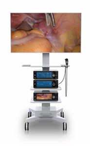
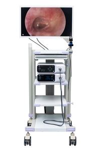
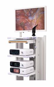
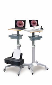
Instruments using fiberoptics and endoscopic remote manipulation, combined with advances in technique, have resulted in consistently good results in the hands of appropriately trained surgeons. Reticulating endoscopes and high-definition cameras offer new visualization capabilities. These advances continue to bolster enthusiasm for these endoscopic approaches.
Transaxillary endoscopic augmentation mammaplasty
The transaxillary approach to breast augmentation was described by Troques in 1972 and Hoehler in 1973. Besides the obvious advantage of the hidden incision, this approach facilitated direct access to the subpectoral plane. With this technique, the inframammary crease was altered and the origin of the pectoralis muscle was dissected blindly, accounting for a significantly higher incidence of implant malposition. The limited exposure of the blind technique did not allow complete division of the prepectoral fascia, resulting in the tendency of high-riding implants or the double-bubble appearance of the inframammary crease.
The advent of endoscopic plastic surgery in the 1990s allowed the application of the endoscope to breast surgery. The Emory group reported their experience with endoscopic breast augmentation through an axillary incision in 1993 using a specialized retractor and an air-filled optical cavity. Ho reported a technique that used glycine irrigation to create a liquid-filled optical cavity, although he now also uses a specialized retractor and an air-filled optical cavity. The increased control resulting from direct visualization of the dissection obviated many of the previous downfalls of the blind axillary approach. Howard demonstrated the benefits of the endoscope with the axillary approach by decreasing the incidence of implant malposition from 8.6% to 2% when the endoscope was used.
Endoscopic transaxillary augmentation mammaplasty is now a widely used technique and has withstood the test of time. However, the learning curve is significant, and more straightforward cases should be considered during the initial experience. The axillary approach has limited application in secondary cases.
Transumbilical endoscopic augmentation mammaplasty (TUBA)
The transumbilical approach was first implemented in 1991 by Johnson and Christ and described in detail in 1993. The technique is unique as it does not employ a local or regional incision but rather uses a remote incision in the umbilicus. The TUBA endured significant early criticism but has gained popularity as it has been shown to be safe and reliable. Though TUBA is technically more challenging, a growing number of plastic surgeons are gaining expertise with this procedure. A primary criticism has been a lack of control of the operative site, especially with regard to bleeding and plane of dissection. With improved instrumentation and a general improvement in endoscopic skills, these criticisms have been proven invalid. The original study by Johnson reported a lower complication rate with less bleeding than other methods.
Relevant Anatomy
Breast shape varies among patients, but knowing and understanding the anatomy of the breast ensures safe surgical planning. When the breasts are carefully examined, significant asymmetries are revealed in most patients. Any preexisting asymmetries, spinal curvature, or chest wall deformities must be recognized and demonstrated to the patient, as these may be difficult to correct and can become noticeable in the postoperative period. Preoperative photographs with multiple views are obtained on all patients and maintained as part of the office record.
The base of the breast overlies the pectoralis major muscle between the second and sixth ribs in the nonptotic state. The gland is anchored to the pectoralis major fascia by the suspensory ligaments first described by Astley Cooper in 1840. These ligaments run throughout the breast tissue parenchyma from the deep fascia beneath the breast and attach to the dermis of the skin. Since they are not taut, they allow for the natural motion of the breast. These ligaments relax with age and time, eventually resulting in breast ptosis. The lower pole of the breast is fuller than the upper pole .