Acute Frontal Sinusitis Surgery
Background and Overview
Acute frontal sinusitis (AFrS) is defined as an acute bacterial infection of the frontal sinus cavity. Among all of the paranasal sinuses, acute bacterial infections localized to the frontal sinus are most commonly associated with intracranial complications. Frontal sinus surgery is performed to prevent potentially life-threatening complications when the infection is unresponsive to maximal medical therapy.
Causes of acute frontal sinusitis
Mucociliary clearance in the frontal sinus occurs in a counterclockwise direction in the right sinus and in a clockwise direction in the left. Secretions are transported along the septal wall to the sinus roof and from there, laterally along the roof and then medially along the floor to reach the ostium. Secretions that are retained because of obstruction serve as a nidus and as growth media for infections.
Given the close anatomic relationship of the ethmoid and frontal sinuses, obstruction of the ethmoid air cells often leads to AFrS. This obstruction may be caused by nasal polyps, tumor, severe septal deviation, trauma, chronic mucosal inflammation, or acute infection. Obstruction impedes the drainage of the frontal and ethmoid sinuses via the frontal recess and impairs mucociliary function.
In addition, there are a number of anatomic variants that lead to obstruction of the nasofrontal outflow tract. These include large concha bullosa, a laterally rotated uncinate process that contacts the middle turbinate, and, conversely, a medially convex middle turbinate that contacts the lateral nasal wall. Previous middle turbinate resection can also lead to stenosis of the frontal sinus ostium secondary to soft-tissue scarring or residual bony fragments.
Clinical presentation
Patients with AFrS often present with dull or pressure-like pain localized to the frontal, supraorbital, or interorbital regions. When this symptom is accompanied by nasal congestion, maxillary pain or pressure, and mucopurulent nasal drainage, the frontal sinus is not likely to be involved in isolation. Isolated AFrS is an important clinical entity, as it suggests a more pernicious type of obstruction of the frontal sinus drainage pathway and may be more liable to result in intracranial or orbital complications.
Complications of acute frontal sinusitis
The complications of AFrS may be classified as either intracranial or orbital. The true incidence of complications stemming from AFrS is not known. Meningitis,brain abscess, Meningitis,brain abscess,
epidural empyema, subdural empyema, cerebral empyema, and dural sinus thrombosis are known intracranial complications of AFrS.
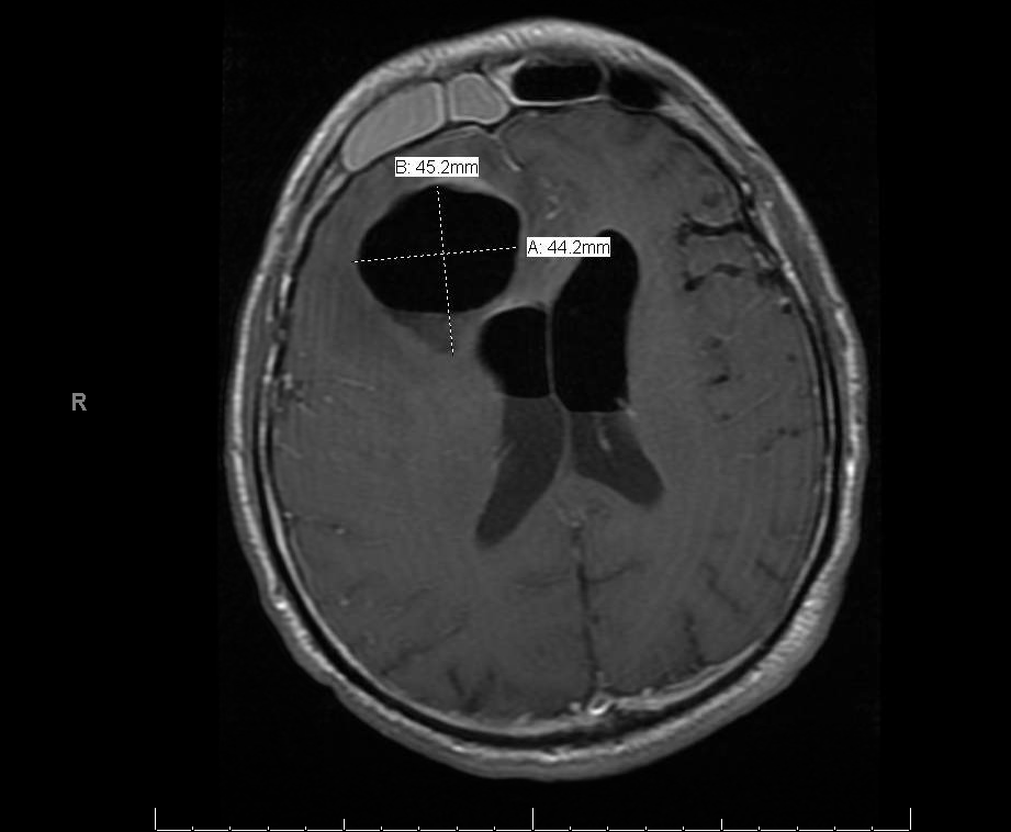
Axial MRI image of a patient with acute frontal sinusitis complicated by large right frontal lobe abscess.
Orbital complications include preseptal or orbital cellulitis, subperiosteal abscess, and cavernous
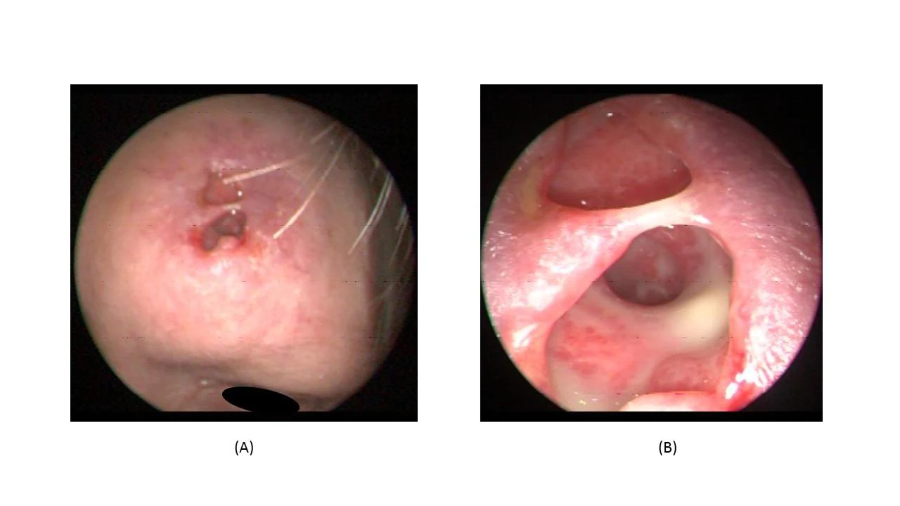
sinus thrombosis.A Pott puffy tumor is a subperiosteal abscess with soft-tissue swelling and pitting edema over the frontal bone. This type of complication results from acute infection of the diploic vein, causing thrombophlebitis. In cases of frontal bone osteomyelitis, a sinocutaneous fistula may result. A few of these complications are relative contraindications to endoscopic sinus surgery and will be discussed later.
A retrospective study by Stokken et al indicated that among pediatric patients who require surgery for complications of acute bacterial sinusitis, the ethmoid and frontal sinuses are more likely to be involved than they are in children undergoing surgery for chronic rhinosinusitis. The study, which involved 27 patients with acute bacterial sinusitis complications and 77 patients with chronic rhinosinusitis, also found that in the former group, there was a lower likelihood of seasonal allergies, prior sinusitis, previous nasal steroid use, or adenoidectomy compared with the chronic rhinosinusitis patients.
History of frontal sinus surgery
Trephination of the frontal sinus has been performed since prehistoric times, either by scraping or incision. Two Peruvian skulls at the Museum of Man, in San Diego, show trephines, with evidence of patient survival. In addition, early surgeries were described in which part of the anterior frontal sinus wall was removed, leaving the patient with a significant cosmetic deformity. More refined surgery of the frontal sinus was first described in the 19th century, and options for treatment have expanded since the advent of endoscopic sinus surgery.
Despite the long history of AFrS surgery, much remains to be elucidated about its long-term outcomes, as well as about postoperative care and follow-up.

